Arteries In Neck Labeled / Anterior Triangle Of The Neck : Internal thoracic artery branches from same segment, but inferiorily, and is therefore not visible.
Arteries In Neck Labeled / Anterior Triangle Of The Neck : Internal thoracic artery branches from same segment, but inferiorily, and is therefore not visible.. Most cases resolve without sequelae with early and appropriate treatment. Superior boundary (base) the lower border of the body of the mandible, and a line extending from the angle of the mandible to the mastoid process Head and neck anatomy is important when considering pathology affecting the same area. The anterior margin of sternocleidomastoid: Internal thoracic artery branches from same segment, but inferiorily, and is therefore not visible.
Head and neck anatomy is important when considering pathology affecting the same area. The anterior margin of sternocleidomastoid: Superior boundary (base) the lower border of the body of the mandible, and a line extending from the angle of the mandible to the mastoid process Superficial dissection of the right side of the neck, showing the carotid and subclavian arteries. Most cases resolve without sequelae with early and appropriate treatment.
Module 22 Muscles And Triangles Of The Neck Anatomy 337 Ereader from wisc.pb.unizin.org Internal thoracic artery branches from same segment, but inferiorily, and is therefore not visible. Branch of vertebral artery and thyrocervical trunk is labeled. Head and neck anatomy is important when considering pathology affecting the same area. Midline of the neck from chin to the jugular notch posterior boundary: Superior boundary (base) the lower border of the body of the mandible, and a line extending from the angle of the mandible to the mastoid process The anterior margin of sternocleidomastoid: Most cases resolve without sequelae with early and appropriate treatment. The common carotid arteries (c) are normal but the right internal jugular vein (j) is enlarged with a dense or enhancing wall surrounding the more lucent intraluminal clot (arrow).
Most cases resolve without sequelae with early and appropriate treatment.
Internal thoracic artery branches from same segment, but inferiorily, and is therefore not visible. In radiology, the 'head and neck' refers to all the anatomical structures in this region excluding the central nervous system, that is, the brain and spinal co. Branch of vertebral artery and thyrocervical trunk is labeled. The common carotid arteries (c) are normal but the right internal jugular vein (j) is enlarged with a dense or enhancing wall surrounding the more lucent intraluminal clot (arrow). Superficial dissection of the right side of the neck, showing the carotid and subclavian arteries. Superior boundary (base) the lower border of the body of the mandible, and a line extending from the angle of the mandible to the mastoid process Midline of the neck from chin to the jugular notch posterior boundary: Most cases resolve without sequelae with early and appropriate treatment. The anterior margin of sternocleidomastoid: Head and neck anatomy is important when considering pathology affecting the same area.
Superior boundary (base) the lower border of the body of the mandible, and a line extending from the angle of the mandible to the mastoid process The common carotid arteries (c) are normal but the right internal jugular vein (j) is enlarged with a dense or enhancing wall surrounding the more lucent intraluminal clot (arrow). Branch of vertebral artery and thyrocervical trunk is labeled. Superficial dissection of the right side of the neck, showing the carotid and subclavian arteries. Internal thoracic artery branches from same segment, but inferiorily, and is therefore not visible.
Major Arteries Of The Head And Neck Carotid Teachmeanatomy from teachmeanatomy.info The common carotid arteries (c) are normal but the right internal jugular vein (j) is enlarged with a dense or enhancing wall surrounding the more lucent intraluminal clot (arrow). Superficial dissection of the right side of the neck, showing the carotid and subclavian arteries. Superior boundary (base) the lower border of the body of the mandible, and a line extending from the angle of the mandible to the mastoid process Most cases resolve without sequelae with early and appropriate treatment. Branch of vertebral artery and thyrocervical trunk is labeled. Internal thoracic artery branches from same segment, but inferiorily, and is therefore not visible. In radiology, the 'head and neck' refers to all the anatomical structures in this region excluding the central nervous system, that is, the brain and spinal co. The anterior margin of sternocleidomastoid:
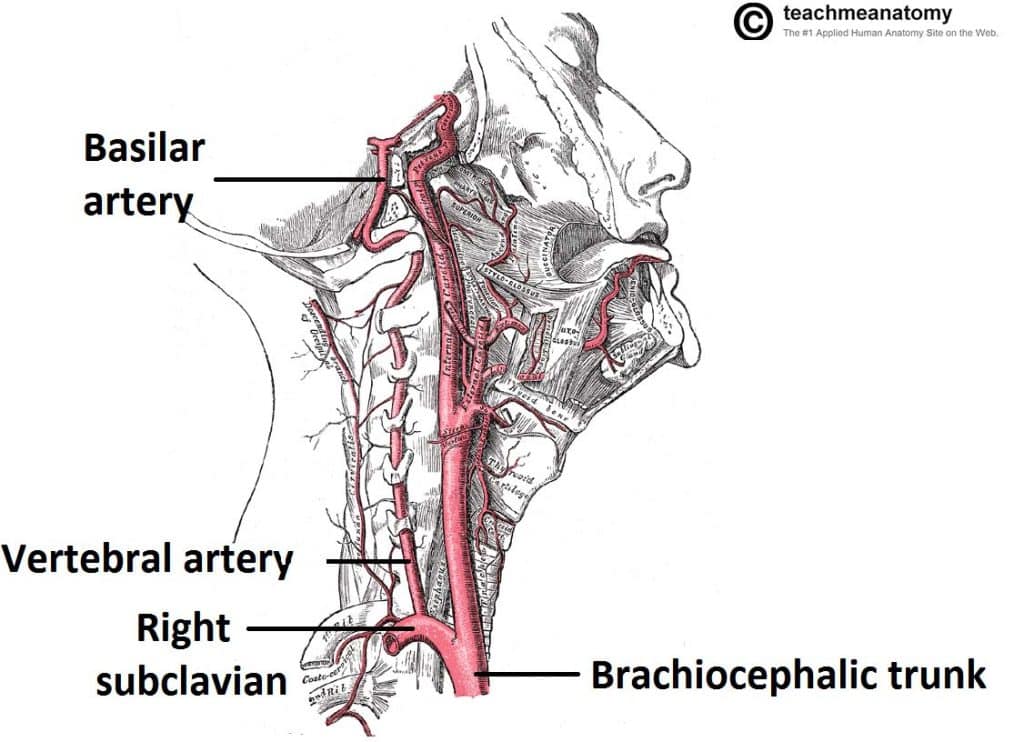
Branch of vertebral artery and thyrocervical trunk is labeled.
Midline of the neck from chin to the jugular notch posterior boundary: Head and neck anatomy is important when considering pathology affecting the same area. Superior boundary (base) the lower border of the body of the mandible, and a line extending from the angle of the mandible to the mastoid process The common carotid arteries (c) are normal but the right internal jugular vein (j) is enlarged with a dense or enhancing wall surrounding the more lucent intraluminal clot (arrow). Superficial dissection of the right side of the neck, showing the carotid and subclavian arteries. Branch of vertebral artery and thyrocervical trunk is labeled. The anterior margin of sternocleidomastoid: Most cases resolve without sequelae with early and appropriate treatment. In radiology, the 'head and neck' refers to all the anatomical structures in this region excluding the central nervous system, that is, the brain and spinal co. Internal thoracic artery branches from same segment, but inferiorily, and is therefore not visible.
Branch of vertebral artery and thyrocervical trunk is labeled. Most cases resolve without sequelae with early and appropriate treatment. The anterior margin of sternocleidomastoid: Internal thoracic artery branches from same segment, but inferiorily, and is therefore not visible. Head and neck anatomy is important when considering pathology affecting the same area.
Head And Neck Arterial System Artwork Stock Image C021 1862 Science Photo Library from media.sciencephoto.com Branch of vertebral artery and thyrocervical trunk is labeled. Midline of the neck from chin to the jugular notch posterior boundary: The common carotid arteries (c) are normal but the right internal jugular vein (j) is enlarged with a dense or enhancing wall surrounding the more lucent intraluminal clot (arrow). Most cases resolve without sequelae with early and appropriate treatment. Head and neck anatomy is important when considering pathology affecting the same area. The anterior margin of sternocleidomastoid: Internal thoracic artery branches from same segment, but inferiorily, and is therefore not visible. Superior boundary (base) the lower border of the body of the mandible, and a line extending from the angle of the mandible to the mastoid process
The anterior margin of sternocleidomastoid:
Head and neck anatomy is important when considering pathology affecting the same area. Branch of vertebral artery and thyrocervical trunk is labeled. Superficial dissection of the right side of the neck, showing the carotid and subclavian arteries. Superior boundary (base) the lower border of the body of the mandible, and a line extending from the angle of the mandible to the mastoid process Most cases resolve without sequelae with early and appropriate treatment. In radiology, the 'head and neck' refers to all the anatomical structures in this region excluding the central nervous system, that is, the brain and spinal co. The common carotid arteries (c) are normal but the right internal jugular vein (j) is enlarged with a dense or enhancing wall surrounding the more lucent intraluminal clot (arrow). The anterior margin of sternocleidomastoid: Midline of the neck from chin to the jugular notch posterior boundary: Internal thoracic artery branches from same segment, but inferiorily, and is therefore not visible.
In radiology, the 'head and neck' refers to all the anatomical structures in this region excluding the central nervous system, that is, the brain and spinal co arteries in neck. Superficial dissection of the right side of the neck, showing the carotid and subclavian arteries.


0 Komentar